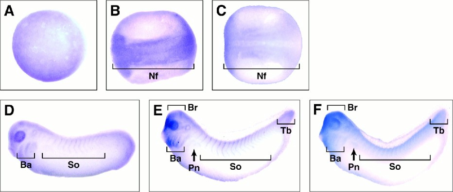
Figure 2. Whole-mount in situ hybridization analysis of XSNT1 and XFGFR1 during embryogenesis. A: Animal pole view of XSNT1 expression in a stage 12 embryo (gastrulation). B: Dorsal view of XSNT1 expression in a stage 19 embryo (neurulation). C: Dorsal view of XFGFR1 expression in a stage 19 embryo. D: Lateral view of XSNT1 expression in stage 26 embryo. E: Lateral view of XSNT1 expression in stage 30 embryo. F: Lateral view of XFGFR1 expression in stage 30 embryo. Nf, neural folds; Ba, branchial arches; So, somites; Br, fore-, mid-, and hindbrain; Pn, pronephros; Tb, tail bud
Image published in: Akagi K et al. (2002)
Copyright © 2002. Image reproduced with permission of the Publisher, John Wiley & Sons.
| Gene | Synonyms | Species | Stage(s) | Tissue |
|---|---|---|---|---|
| frs2.L | frs2a, frs2alpha, snt, snt-1, snt1, xfrs2, XSNT, xsrf2 | X. laevis | Throughout NF stage 12 | animal hemisphere |
| fgfr1.S | bfgfr, cek, fgfr-1, fgfr1-a, fgfr1-b, flg, flt-2, kal2, ogd, X1FGFR, XFGFR-1, xfgfr1, xfgfra2 | X. laevis | Throughout NF stage 17 | neural plate anterior neural fold chordal neural plate pre-chordal neural plate neuroectoderm |
| frs2.L | frs2a, frs2alpha, snt, snt-1, snt1, xfrs2, XSNT, xsrf2 | X. laevis | Throughout NF stage 19 | anterior neural fold neural plate neural groove neuroectoderm |
| frs2.L | frs2a, frs2alpha, snt, snt-1, snt1, xfrs2, XSNT, xsrf2 | X. laevis | Throughout NF stage 26 | forebrain midbrain hindbrain somite central nervous system brain spinal cord otic placode eye cranial neural crest mandibular crest hyoid crest anterior branchial crest posterior branchial crest trunk somite tail bud presomitic mesoderm |
| frs2.L | frs2a, frs2alpha, snt, snt-1, snt1, xfrs2, XSNT, xsrf2 | X. laevis | Throughout NF stage 29 and 30 | forebrain hindbrain midbrain eye primordium somite branchial arch otic vesicle tail bud brain spinal cord pharyngeal arch mandibular arch hyoid arch eye olfactory region midbrain-hindbrain boundary anterior placodal area |
| fgfr1.S | bfgfr, cek, fgfr-1, fgfr1-a, fgfr1-b, flg, flt-2, kal2, ogd, X1FGFR, XFGFR-1, xfgfr1, xfgfra2 | X. laevis | Throughout NF stage 29 and 30 | otic vesicle pronephric kidney somite tail bud eye primordium branchial arch forebrain hindbrain midbrain pharyngeal arch mandibular arch hyoid arch |
Image source: Published
Permanent Image Page
Printer Friendly View
XB-IMG-42563