Click here to close
Hello! We notice that you are using Internet Explorer, which is not supported by Xenbase and may cause the site to display incorrectly.
We suggest using a current version of Chrome,
FireFox, or Safari.
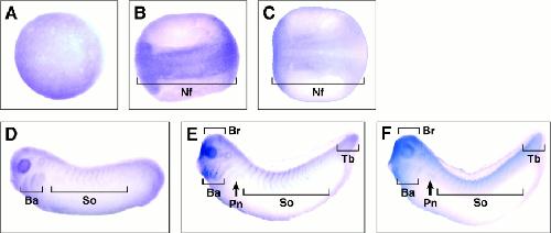
Figure 2. Whole-mount in situ hybridization analysis of XSNT1 and XFGFR1 during embryogenesis. A: Animal pole view of XSNT1 expression in a stage 12 embryo (gastrulation). B: Dorsal view of XSNT1 expression in a stage 19 embryo (neurulation). C: Dorsal view of XFGFR1 expression in a stage 19 embryo. D: Lateral view of XSNT1 expression in stage 26 embryo. E: Lateral view of XSNT1 expression in stage 30 embryo. F: Lateral view of XFGFR1 expression in stage 30 embryo. Nf, neural folds; Ba, branchial arches; So, somites; Br, fore-, mid-, and hindbrain; Pn, pronephros; Tb, tail bud