
ECB-IMG-208673
Echinobase Image ID: 208673
|
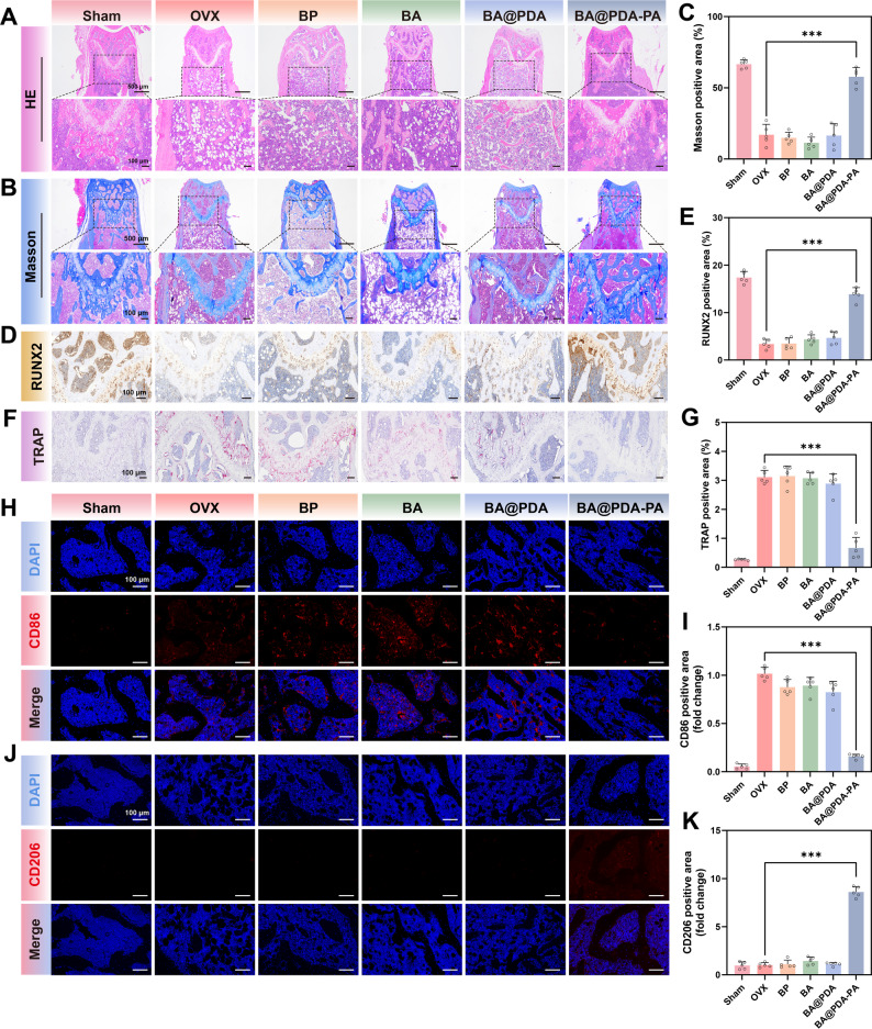
Fig. 8. Histological staining of mice femora. (A) HE staining of the distal femur of mice. (B) Masson staining of the distal femur of mice. (C) Quantitative results of the ratio of the new bone area to the total bone area in Masson staining. (D) Immunohistochemistry staining of RUNX2. (E) Quantitative analysis of RUNX2 positive area in immunohistochemistry staining. (F) TRAP staining of the distal femur of mice. (G) Quantitative results of the TRAP positive area to the total bone area in TRAP staining. (H) Immunofluorescence staining images of CD86 in the distal femur of mice. (I) Quantitative analysis of CD86 in immunofluorescence staining. (J) Immunofluorescence staining images of CD206 in the distal femur of mice. (K) Quantitative analysis of CD206 in immunofluorescence staining. Data are means ± S.D. (n = 5), ***p < 0.001 Image published in: Shen X et al. (2026) Image downloaded from an Open Access article in PubMed Central. © The Author(s) 2026 Larger Image Printer Friendly View |