
ECB-IMG-208623
Echinobase Image ID: 208623
|
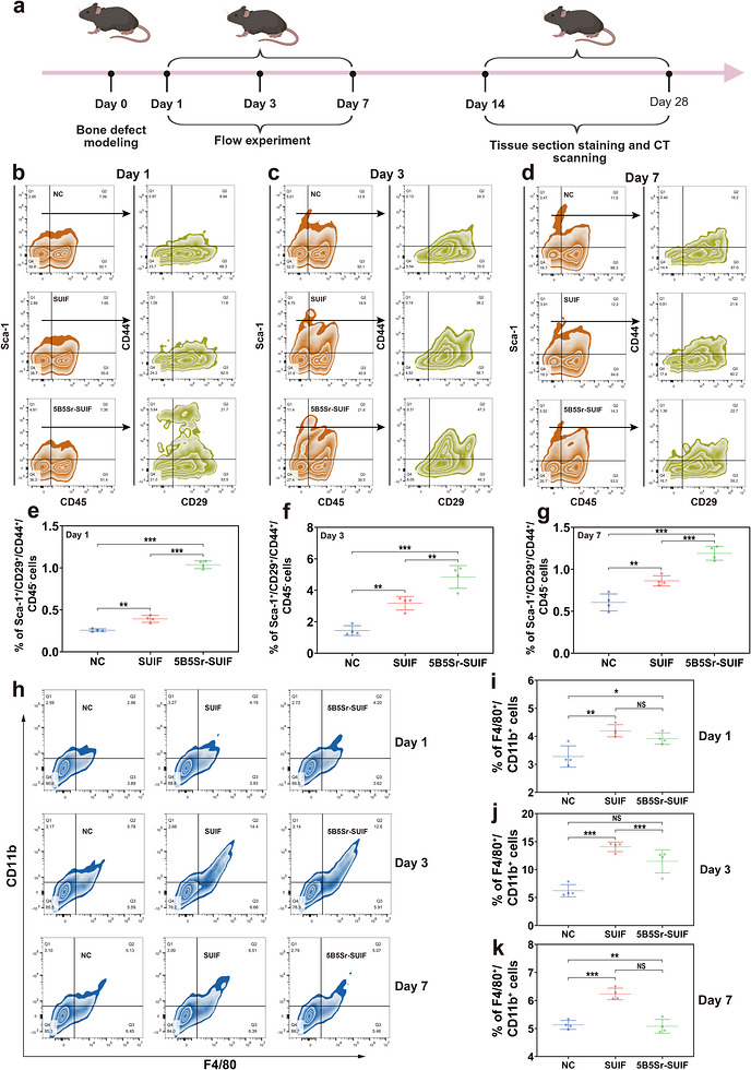
FIGURE 5. Material‐mediated recruitment of mesenchymal stem cells and macrophages in vivo. (a) Schematic timeline of tissue harvest at different time points after material implantation for subsequent analyses. Flow cytometric analysis of MSC‐related surface markers Sca‐1, CD29, CD44, and the exclusion marker CD45 in peri‐defect tissue cells from the blank control (NC), SUIF, and 5B5Sr‐SUIF groups at postoperative day 1 (b), day 3 (c), and day 7 (d). Quantitative analysis of the proportion of MSC‐characteristic cell populations (Sca‐1+/CD29+/CD44+/CD45−) at day 1 (e), day 3 (f), and day 7 (g) post‐surgery. (h) Flow cytometric detection of macrophage‐characteristic surface markers CD11b and F4/80 in peri‐defect tissue cells from the three groups at postoperative days 1, 3, and 7. Quantitative analysis of the proportion of macrophage‐characteristic cell populations (F4/80+/CD11b+) at day 1 (i), day 3 (j), and day 7 (k) post‐surgery. Image published in: Lu Y et al. (2026) Image downloaded from an Open Access article in PubMed Central. © 2026 The Author(s). Endocrinology, Diabetes & Metabolism published by John Wiley & Sons Ltd. Larger Image Printer Friendly View |