
ECB-IMG-208622
Echinobase Image ID: 208622
|
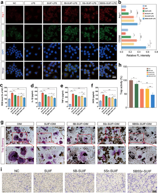
FIGURE 4. In vitro MSC–macrophage communication driven by the SUIF platform. (a) Immunofluorescence staining of ARG‐1 and CD206 in macrophages after 48 h of treatment in the material–MSCs–macrophage co‐culture system. (b) Quantitative analysis of ARG‐1 and CD206 fluorescence intensity. (c) Assessment of intracellular ROS levels in macrophages after 48 h of treatment in the material‐MSCs‐macrophage co‐culture system. (d–f) Evaluation of pro‐inflammatory mediators IL‐1β (d), TNF‐α (e), and iNOS (f) in the material‐MSCs‐macrophage co‐culture system after 48 h of treatment. (g) The TRAP staining outcomes for the OIM group, SUIF+OIM group, 5B‐SUIF+OIM group, 5Sr‐SUIF+OIM group, and 5B5Sr‐SUIF+OIM group following stimulation with osteoclast induction medium (OIM) in the material–MSCs–macrophage co‐culture system. (h) Quantitative analysis of osteoclastic activity. (i) Transwell assay for the in vitro recruitment of MSCs. Image published in: Lu Y et al. (2026) Image downloaded from an Open Access article in PubMed Central. © 2026 The Author(s). Endocrinology, Diabetes & Metabolism published by John Wiley & Sons Ltd. Larger Image Printer Friendly View |