
ECB-IMG-208592
Echinobase Image ID: 208592
|
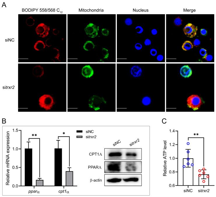
Figure 5. PLIN2-TRXR2 module promotes lipid droplets-to-mitochondria fatty acid flux. (A) Coelomocytes were transfected with siNC or sitrxr2 for 24 h, followed by treatment with 500 μM oleic acid (OA) for 12 h. Cells were then incubated with BODIPY 558/568 C12 (red) for 30 min (pulse) to label lipid droplet-associated fatty acids. After washing with PBS, cells were incubated in Leibovitz’s L-15 medium for 6 h (chase). Cells were then fixed and stained with Mito-Tracker Green and DAPI (blue). Images were acquired by confocal microscopy. Scale bar = 5 μM (B,C) Coelomocytes were transfected with siNC or sitrxr2 for 24 h, followed by treatment with 500 μM OA for 12 h. Cells were then incubated in Leibovitz’s L-15 medium for an additional 6 h. Relative mRNA and protein expression of PPARA and CPT1A (B) was detected by qRT-PCR and WB (B). Intracellular ATP content was determined according to the manufacturer’s protocol (C). Data from at least three independent biological replicates (mean ± SD) or representative data. Significantly different experimental groups: * p < 0.05, ** p < 0.01 by Student’s t test. CPT1A, O-palmitoyltransferase 1 (CPT1A); PPARA, peroxisome proliferator-activated receptor alpha; PLIN2, perilipin 2; TRXR2, thioredoxin reductase 2, mitochondrial. Image published in: Fan H et al. (2026) Image downloaded from an Open Access article in PubMed Central. © 2026 by the authors. Larger Image Printer Friendly View |