
ECB-IMG-208589
Echinobase Image ID: 208589
|
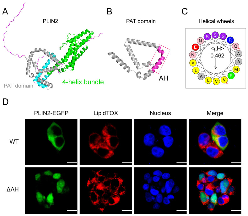
Figure 2. PLIN2 binds lipid droplets via the PAT domain. (A) Predicted three-dimensional structure of PLIN2. The N-terminal PAT domain is highlighted in gray, and the C-terminal region forms a four-helix bundle (shown in green). (B) Enlarged view of the PAT domain, with the predicted amphipathic helix (AH) highlighted in magenta (dashed box). (C) Helical wheel projection of the predicted AH of PLIN2 generated using the HeliQuest tool. The hydrophobic moment (μH) represents the amphipathic nature of the helix. (D) Subcellular localization of PLIN2 and the AH-deficient variant (ΔAH). HEK293T cells were transfected with PLIN2-EGFP (WT) or ΔAH-EGFP (ΔAH) (green) for 24 h, followed by fixation and staining with LipidTOX (red) to label lipid droplets and DAPI (blue) for nucleus. Images were acquired by confocal microscopy. Scale bar = 10 μM. PLIN2, perilipin 2. Image published in: Fan H et al. (2026) Image downloaded from an Open Access article in PubMed Central. © 2026 by the authors. Larger Image Printer Friendly View |