
ECB-IMG-180266
Echinobase Image ID: 180266
|
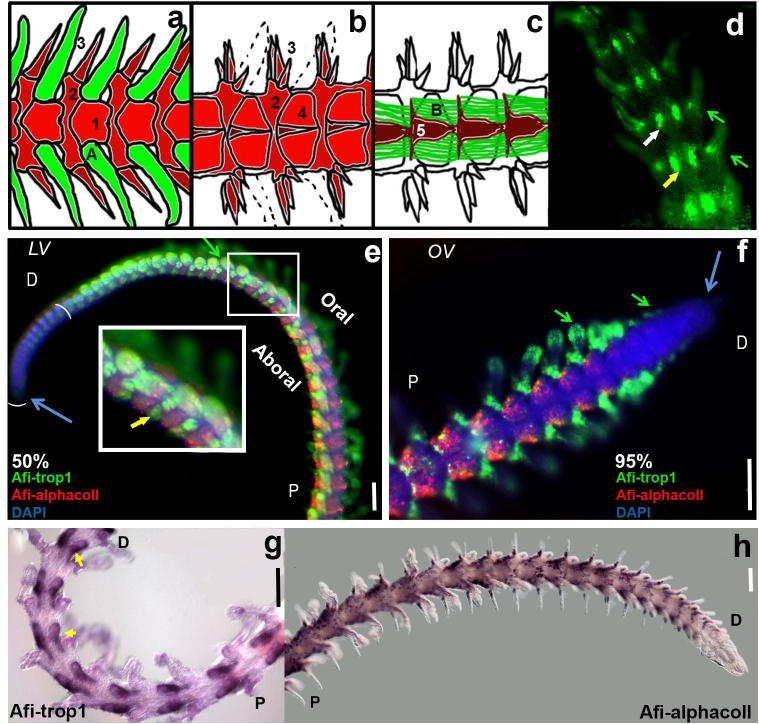
Fig. 1. (aâd) Schematic diagram of muscle and skeletal structures within the brittle star arm. (a) Oral view showing the spines, podia, and lateral and oral arm shields. (b) Aboral view showing spines, and lateral and aboral arm shields. (c) View of internal structures including the vertebrae and the intervertebral muscles. (d) Phalloidin staining revealing muscle structures in the proximal part of the 95% regenerating arm (aboral view). (e, f) Double fluorescent in situ hybridization showing Afi-trop1 localized to the podia (green arrows) at the oral side and intervertebral muscles (yellow arrow) at the aboral side of the 50% (e) and 95% (f) regenerating arm. Afi-αcoll is restricted to lateral arm shields and the base of the spines. The two differentiation markers are not co-expressed and neither is found at the distal tip (blue arrow) of the regenerating arm which confirms their use as markers for specified structures. (g,h) Chromogenic single in situ hybridization clearly showing expression in 95% regenerating arms of Afi-trop1 in the intervertebral muscles (yellow arrows) as well as the podia and Afi-αcoll in the lateral shields and base of the spines. Scale bars â 100 μm. Green â muscle structures, red â skeletal structures, 1 â oral arm shield, 2 â lateral arm shield, 3 â spines, 4 â aboral arm shields, 5 â vertebrae, A â podia, B â intervertebral muscles. Green arrows â podia, yellow arrows â intervertebral muscles, blue arrows â distal tip. OV â oral view, LV â lateral view. P â proximal, D â distal. Image published in: Czarkwiani A et al. (2013) Image downloaded from an Open Access article in PubMed Central. © 2013 The Authors Larger Image Printer Friendly View |