
ECB-IMG-177887
Echinobase Image ID: 177887
|
|
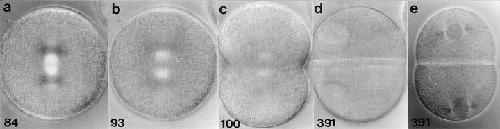
Figure 6. The development of a zygote treated with protein synthesis inhibitors beginning at first prophase. (a) Prometaphase. (b) Anaphase. (c) Initiation of cytokinesis at telophase. (d) The blastomeres arrest in interphase of the second cell cycle, and the nuclei become enlarged over time. The two asters in each blastomere are weakly birefringent and thus not visible in this micrograph. (e) Another zygote from the same culture, also arrested in second interphase. This zygote has been treated with 2% hexylene glycol to augment astral birefringence. Minutes after fertilization are shown in the lower corner of each panel. Polarization optics. 10 μm per scale division. Image published in: Hinchcliffe EH et al. (1998) Image downloaded from an Open Access article in PubMed Central. Image reproduced on Echinobase with permission of the publisher and the copyright holder. Larger Image Printer Friendly View |