|
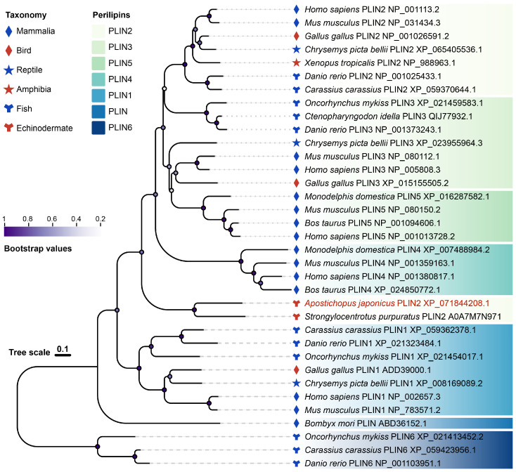
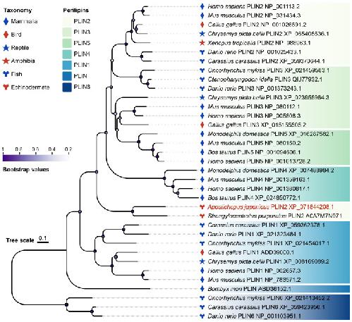
Figure 1. Phylogenetic analysis of perilipin (PLIN) family proteins across representative species. A maximum-likelihood phylogenetic tree was constructed using amino acid sequences of PLIN family members from vertebrates and invertebrates. Different PLIN family members (PLIN1-PLIN6) are indicated by distinct color blocks on the right. Major taxonomic groups are denoted by symbols, including mammals, birds, reptiles, amphibians, fish, and echinoderms. Apostichopus japonicus PLIN2 is highlighted in red. Node support is represented by a color gradient corresponding to bootstrap values. The scale bar represents the number of amino acid substitutions per site. |
|
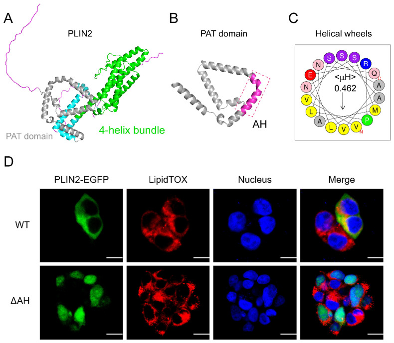
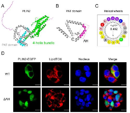
Figure 2. PLIN2 binds lipid droplets via the PAT domain. (A) Predicted three-dimensional structure of PLIN2. The N-terminal PAT domain is highlighted in gray, and the C-terminal region forms a four-helix bundle (shown in green). (B) Enlarged view of the PAT domain, with the predicted amphipathic helix (AH) highlighted in magenta (dashed box). (C) Helical wheel projection of the predicted AH of PLIN2 generated using the HeliQuest tool. The hydrophobic moment (μH) represents the amphipathic nature of the helix. (D) Subcellular localization of PLIN2 and the AH-deficient variant (ΔAH). HEK293T cells were transfected with PLIN2-EGFP (WT) or ΔAH-EGFP (ΔAH) (green) for 24 h, followed by fixation and staining with LipidTOX (red) to label lipid droplets and DAPI (blue) for nucleus. Images were acquired by confocal microscopy. Scale bar = 10 μM. PLIN2, perilipin 2. |
|
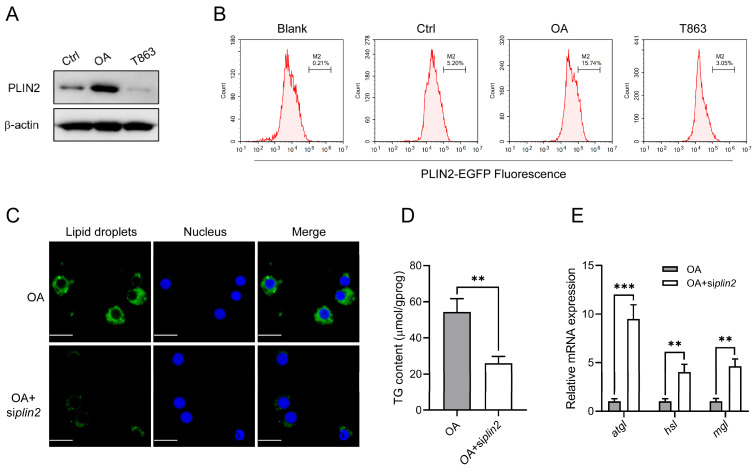
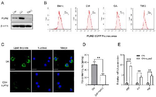
Figure 3. PLIN2 stabilizes lipid droplets by restraining lipolytic activation. (A) Coelomocytes were treated with OA or T863 for 12 h. Protein expression of PLIN2 was determined by Western blot, with β-actin as the loading control. Data represent three independent experiments. (B) HEK293T cells were transfected with the PLIN2-EGFP expression plasmid. After 24 h, cells were treated with OA or T863 for 12 h, harvested, and analyzed by flow cytometry using the FITC channel to quantify EGFP fluorescence intensity. (C–E) Coelomocytes were transfected with siNC or siplin2. After 24 h, cells were incubated with 500 μM OA for 12 h, followed by fixation and staining with BODIPY 493/503 (green) to label lipid droplets and DAPI (blue) for nucleus. Images were acquired by confocal microscopy. Scale bar = 10 μM (C). Cellular triglyceride content was measured (D), and relative mRNA expression of atgl, hsl, and mgl was detected by qRT-PCR (E). Data from at least three independent biological replicates (mean ± SD). Significantly different experimental groups: ** p < 0.01, *** p < 0.001 by Student’s t test. atgl, adipose triglyceride lipase; hsl, hormone-sensitive lipase; mgl, monoacylglycerol lipase; PLIN2, perilipin 2; OA, oleic acid. |
|
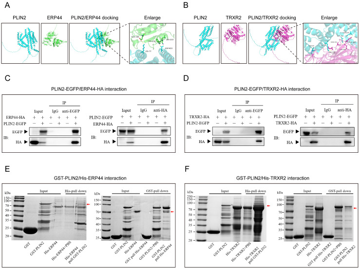
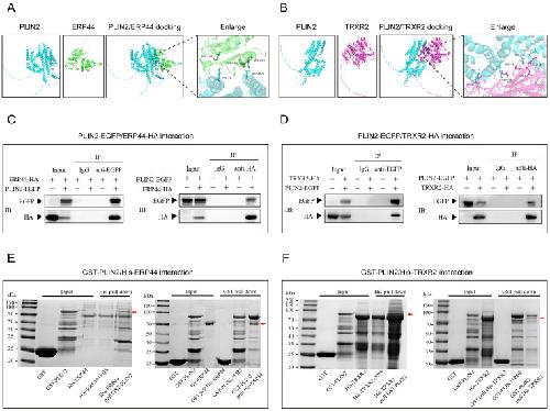
Figure 4. PLIN2 interacts with ERP44 and TRXR2. (A,B) Predicted interactions of PLIN2 with ERP44 and TRXR2 based on AlphaFold3 modeling, visualized using PyMOL2. (C,D) Co-immunoprecipitation validation in HEK293T cells. Cells cultured in 10 cm dishes were co-transfected with PLIN2-EGFP and ERP44-HA or TRXR2-HA plasmids for 48 h. IP was implemented with anti-EGFP or anti-HA beads. IB was conducted with anti-HA and anti-EGFP antibodies, respectively; mouse IgG was used as the control. Data represent three independent biological replicates. (E,F) In vitro pull-down validation. Recombinant GST-PLIN2, His-ERP44, and TRXR2 were expressed in E. coli. His pull-down was performed by incubating His-ERP44 or His-TRXR2 with GST-PLIN2, followed by capture with His-affinity resin. Reciprocal GST pull-down assays were performed using GST-Sepharose resin. Bound proteins were analyzed by SDS-PAGE. The red arrow indicates the protein band pulled down after the interaction. ERP44, endoplasmic reticulum resident protein 44; PLIN2, perilipin 2; TRXR2, thioredoxin reductase 2, mitochondrial. |
|
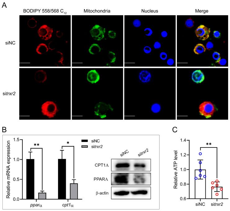
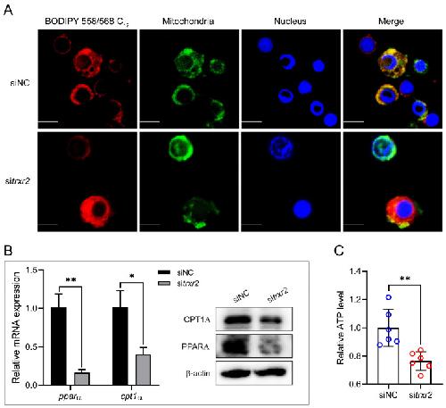
Figure 5. PLIN2-TRXR2 module promotes lipid droplets-to-mitochondria fatty acid flux. (A) Coelomocytes were transfected with siNC or sitrxr2 for 24 h, followed by treatment with 500 μM oleic acid (OA) for 12 h. Cells were then incubated with BODIPY 558/568 C12 (red) for 30 min (pulse) to label lipid droplet-associated fatty acids. After washing with PBS, cells were incubated in Leibovitz’s L-15 medium for 6 h (chase). Cells were then fixed and stained with Mito-Tracker Green and DAPI (blue). Images were acquired by confocal microscopy. Scale bar = 5 μM (B,C) Coelomocytes were transfected with siNC or sitrxr2 for 24 h, followed by treatment with 500 μM OA for 12 h. Cells were then incubated in Leibovitz’s L-15 medium for an additional 6 h. Relative mRNA and protein expression of PPARA and CPT1A (B) was detected by qRT-PCR and WB (B). Intracellular ATP content was determined according to the manufacturer’s protocol (C). Data from at least three independent biological replicates (mean ± SD) or representative data. Significantly different experimental groups: * p < 0.05, ** p < 0.01 by Student’s t test. CPT1A, O-palmitoyltransferase 1 (CPT1A); PPARA, peroxisome proliferator-activated receptor alpha; PLIN2, perilipin 2; TRXR2, thioredoxin reductase 2, mitochondrial. |
|
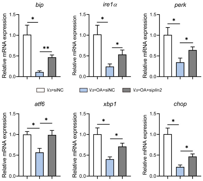
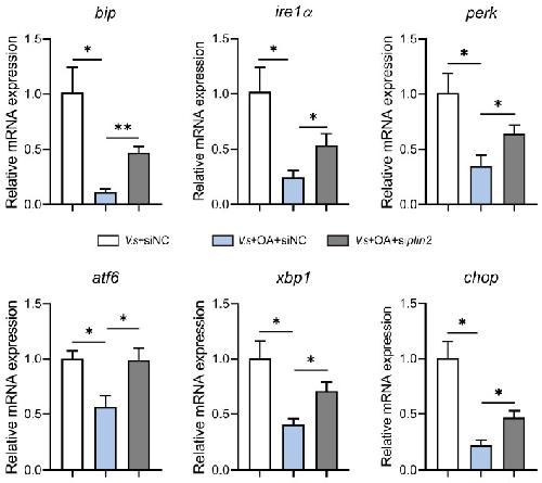
Figure 6. PLIN2-dependent lipid droplet organization mitigates Vibrio splendidus-induced ER stress. Coelomocytes were transfected with siNC or siplin2 for 24 h, followed by infection with V. splendidus (MOI = 10) for 12 h in the absence or presence of 500 μM OA. Relative mRNA expression of bip, ire1α, perk, atf6, xbp1, and chop was detected by qRT-PCR. Data are presented as the mean ± SD from three independent biological replicates. Significantly different experimental groups: * p < 0.05, ** p < 0.01 by one-way ANOVA. atf6, cyclic AMP-dependent transcription factor atf-6 alpha; bip, endoplasmic reticulum chaperone bip; chop, DNA damage-inducible transcript 3 protein; ire1α, serine/threonine-protein kinase/endoribonuclease ire1α; OA, oleic acid; perk, eukaryotic translation initiation factor 2-alpha kinase 3; plin2, perilipin 2; xbp1, x-box-binding protein 1. |
|
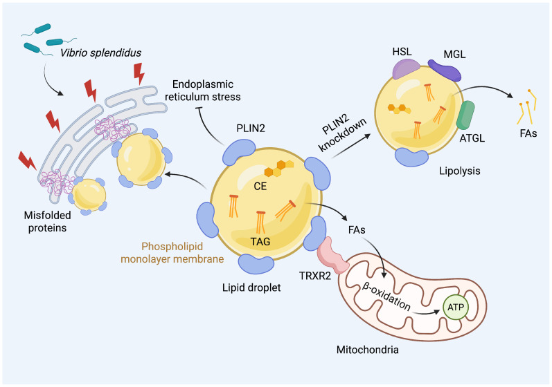
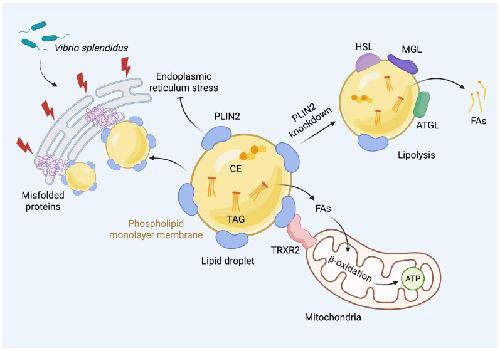
Figure 7. Roles of PLIN2 in coelomocytes of Apostichopus japonicus. PLIN2 localizes to lipid droplets and restricts access of lipases, such as ATGL, MGL, and HSL, to the droplet surface, thereby inhibiting excessive lipolysis. The interaction between PLIN2 and TRXR2 facilitates the transfer of fatty acid from lipid droplets to mitochondria for β-oxidation. Additionally, PLIN2 contributes to the alleviation of Vibrio splendidus-induced ER stress. ATGL, adipose triglyceride lipase; CE, cholesteryl ester; FAs, fatty acids; HSL, hormone-sensitive lipase; MGL, monoacylglycerol lipase; PLIN2, perilipin 2; TAG, triacylglycerol. |