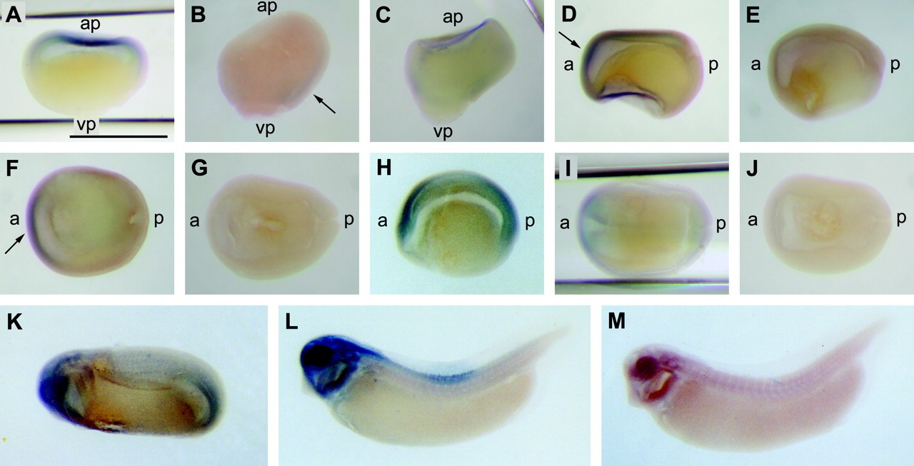
FIG. 6. XMIF is expressed in the developing central nervous system. Whole mount in situ hybridization analysis was performed for the blastula (stage 9), gastrula (stage 11), early neurula (stage 13), neurula (stage 15), early tailbud (stage 26), and tailbud (stage 34) embryos. A, lateral view of a stage 9 embryo hybridized with the MIF probe. No signal was detected in control hybridization using a sense probe (not shown). B and C, lateral views of stage 11 embryos hybridized with antisense (B) and sense (C) probes. The arrow in B indicates the signal in the dorsal marginal zone. D-G, lateral (D and E) and dorsal (F and G) views of stage 13 embryos hybridized with antisense (D and F) and sense (E and G) probes. The arrows in D and F indicate the expression in the anterior region of the neural plate. H-J, lateral (H) and dorsal (I and J) views of stage 15 embryos hybridized with antisense (H and I) and sense (J) probes. K, lateral view of a stage 26 embryo. L and M, lateral views of stage 34 embryos hybridized with antisense (L) and sense (M) probes. The bar shown in A indicates 1 mm. ap, animal pole; vp, vegetal pole; a, anterior; p, posterior.
Image published in: Suzuki M et al. (2004)
Copyright © 2004. Image reproduced with permission of the Publisher.
| Gene | Synonyms | Species | Stage(s) | Tissue |
|---|---|---|---|---|
| mif.L | mmif | X. laevis | Throughout NF stage 11 | animal pole dorsal marginal zone |
| mif.L | mmif | X. laevis | Throughout NF stage 13 | neural plate |
| mif.L | mmif | X. laevis | Throughout NF stage 15 | neural plate neuroectoderm |
| mif.L | mmif | X. laevis | Throughout NF stage 26 | optic vesicle brain forebrain midbrain hindbrain tail region |
| mif.L | mmif | X. laevis | Throughout NF stage 33 and 34 | head region brain forebrain midbrain hindbrain head mesenchyme spinal cord |
| mif.L | mmif | X. laevis | Throughout NF stage 9 | animal pole |
Image source: Published
Permanent Image Page
Printer Friendly View
XB-IMG-87079