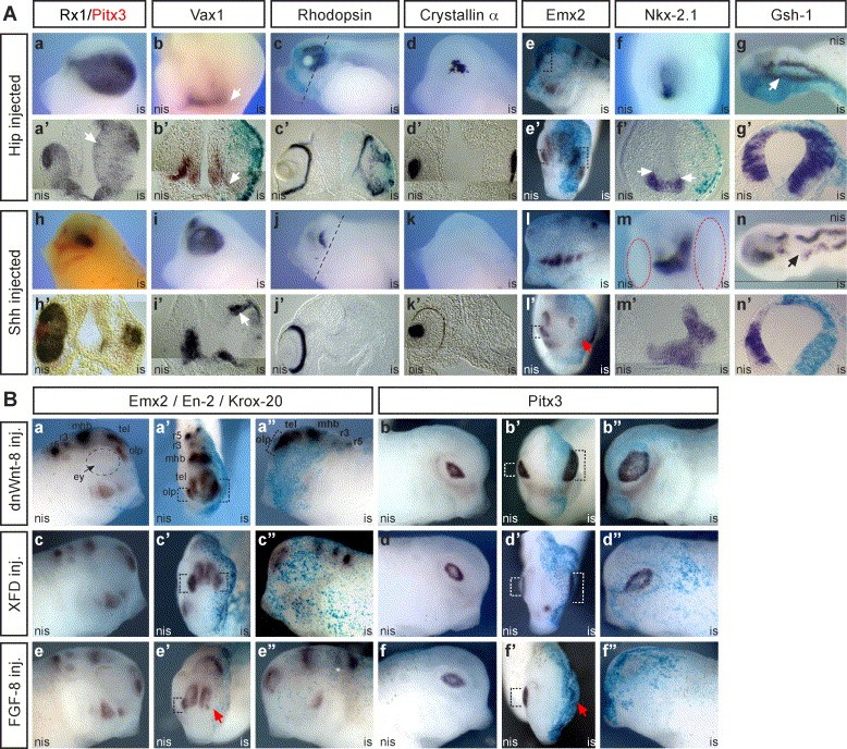
Fig. 5. The inhibition of Shh, Fgf and Wnt-8 signaling affects eye, brain, and placode development in tadpole stage embryos. (A) Overexpression of mHip1 and Shh. (aâgâ²) Microinjection of mHip1 (750 pg) into one cell of two-cell stage embryos. (hânâ²) Overexpression of Shh (500 pg) into one cell of two-cell stage embryos. (b, eâ², f, lâ², m) Frontal view. (a, c, d, e, h, i, j, k, l) Lateral view. (g, n) Dorsal view. (aâ²âdâ², fâ², gâ², hâ²âkâ², mâ², nâ²) Transversal sections. (a, aâ²) Formation of a giant eye. Note Xrx1-positive cells extend into the prospective midbrain region (white arrow in aâ², 67%, n = 18). (b, bâ²) Repression of optic stalk marker Vax1 within the ventral fore- and midbrain (white arrows, 63%, n = 28). (c, câ²) Displaced Rhodopsin positive cells in the enlarged eye (76%, n = 21). (d, dâ²) Abnormal lens formation revealed by Crystallin α expression (50%, n = 12). (e, eâ²) Enlarged olfactory placode as revealed by Emx2 expression, indicated by dashed open rectangles (65%, n = 17). (f, fâ²) Ventral shift of the dorsal limit of Nkx-2.1 expression (white arrows in fâ², 55%, n = 11). (g, gâ²) Ventral expansion of Gsh-1 expression (38%, n = 8). (hâhâ²) Note the reduction of retinal tissue (Xrx1, dark brown) and lens tissue (Pitx3, red, 50%, n = 10). (i, iâ²) Expanded expression domain of the optic stalk marker Vax1 (white arrow in iâ², 62%, n = 13). (j, jâ²) Loss of Rhodopsin positive cells (83%, n = 16). (k, kâ²) Absence of lens specific Crystallin α expression (57%, n = 14). (l, lâ²) Suppression of olfactory placode development as revealed by the loss of Emx2 expression, indicated by a red arrow (65%, n = 17). The dashed open rectangle marks the olfactory placode on the non-injected side (lâ²). (m, mâ²) Dorsal shift of ventral forebrain tissue as revealed by Nkx-2.1 expression (56%, n = 16), the eye structures are emphasized by dashed ellipses. (n, nâ²) Reduced expression of the dorsal neural tube marker Gsh-1 in the prospective midbrain (black arrow, 42%, n = 12). LacZ mRNA was co-injected as a lineage tracer (light blue in bâ², c, câ², e, eâ², fâ², g, gâ², l, lâ², nâ²). (B) Influence of dnWnt-8, XFD and Fgf-8 overexpression on lens and olfactory placode development. (aâ², bâ², câ², dâ², eâ², fâ²) Frontal views, dashed open rectangles indicate the size of the olfactory and lens placodes, respectively. (a, aâ², b, bâ², c, câ², d, dâ², e, eâ², f, fâ²) Lateral views. (aâbâ²) Overexpression of dnWnt-8 (500 pg) resulted in enlarged olfactory (Emx2; 76%, n = 22) and lens placodes (Pitx3; 78%, n = 18). (câdâ²) XFD (1 ng) injection did not alter the size of the olfactory placode but led to enlarged lens placodes (83%, n = 16). (eâfâ²) Additional Fgf-8 (10 pg) inhibits olfactory and lens placode induction (red arrows in eâ² and fâ²; 76%, n = 19). (aâaâ², câcâ², eâeâ²) Krox-20 and En-2 expression was followed to control hindbrain and midbrainâhindbrain formation, respectively. LacZ mRNA was co-injected as a lineage tracer (light blue in aâ², aâ², bâ², bâ², câ², câ², dâ², dâ², eâ², eâ², fâ², fâ²). Abbreviations: ey, eye cup; mhb, midbrainâhindbrain boundary; olp, olfactory placode; r3, rhombomere 3; r5, rhombomere 5; tel, telencephalon.
Image published in: Cornesse Y et al. (2005)
Copyright © 2005. Image reproduced with permission of the Publisher, Elsevier B. V.
Permanent Image Page
Printer Friendly View
XB-IMG-43415