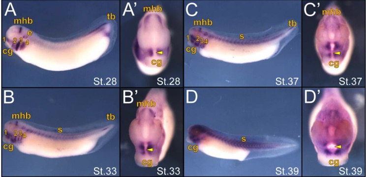
Figure 4: In situ hybridization of ism1 expression in late stage Xenopus laevis embryos. Embryos are shown in a lateral (A-D) or anterior (Aâ-Dâ) view with the cement gland (cg) labeled for ventral orientation. (A) Stage 28 embryo showing strong expression in the branchial arches (ba), midbrain-hindbrain boundary (mhb), ear placode (e), and tailbud (tb) with the yellow arrowheads marking the primitive mouth. (B) Stage 33 embryos showing decreased expression in the branchial arches (ba), midbrain-hindbrain boundary (mhb) and tailbud (tb), with concentrated expression surrounding the primitive mouth (arrowhead) and expression in the somites (s). (C-D) Stage 37 and 39 embryos showing continued concentrated ism1 expression surrounding the primitive mouth (arrowhead).
Image published in: Lansdon LA et al. (2017)
Copyright © 2017. Image reproduced with permission of the Publisher.
| Gene | Synonyms | Species | Stage(s) | Tissue |
|---|---|---|---|---|
| ism1.L | ism-1, isthmin, Isthmin-1, xISM, xISM-1 | X. laevis | Throughout NF stage 28 | midbrain-hindbrain boundary pharyngeal arch mandibular arch hyoid arch branchial arch tail tip otic vesicle mouth primordium |
| ism1.L | ism-1, isthmin, Isthmin-1, xISM, xISM-1 | X. laevis | Throughout NF stage 33 and 34 | midbrain-hindbrain boundary pharyngeal arch mandibular arch hyoid arch branchial arch somite trunk somite tail tip mouth primordium |
| ism1.L | ism-1, isthmin, Isthmin-1, xISM, xISM-1 | X. laevis | Throughout NF stage 37 and 38 | midbrain-hindbrain boundary pharyngeal arch mandibular arch hyoid arch branchial arch tail tip otic vesicle mouth primordium trunk somite |
| ism1.L | ism-1, isthmin, Isthmin-1, xISM, xISM-1 | X. laevis | Throughout NF stage 39 | pharyngeal arch somite trunk somite tail somite head mouth primordium |
Image source: Published
Permanent Image Page
Printer Friendly View
XB-IMG-171272