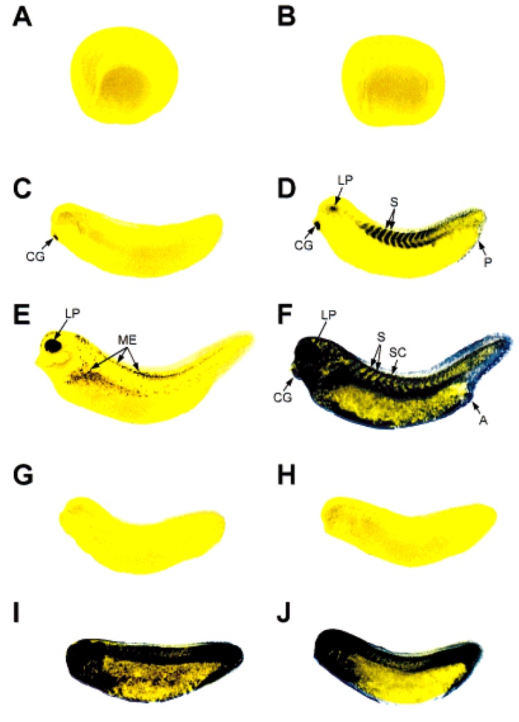
Fig. 2. Spatial pattern of hsp30 mRNA accumulation during early Xenopus development. Whole-mount in situ hybridization with DIGlabeled hsp30 antisense riboprobe was carried out with control (A,C,E) and heat shocked (B,D,F; 1 h at 33°C) Xenopus embryos at neurula (A,B; stage 17/18), mid- (C,D; stage 28) and late (E,F; stage 35) tailbud stages. At the late tailbud stage, control embryos (E) display a natural increase in eye pigmentation and melanocyte production. G,H: Control and heat shocked midtailbud stage embryos (stage 28), respectively, hybridized with DIG-labeled hsp30 sense riboprobe. I,J: Control and heat shocked midtailbud stage embryos (stage 28), respectively, hybridized with DIG-labeled actin antisense riboprobe. LP, lens placode; S, somites; P, proctodeum; CG, cement gland; SC, spinal cord; A, anus; ME, melanocytes.
Image published in: Lang L et al. (1999)
Copyright © 1999. Image reproduced with permission of the Publisher.
| Gene | Synonyms | Species | Stage(s) | Tissue |
|---|---|---|---|---|
| hsp30c.L | X. laevis | Throughout NF stage 17 to NF stage 18 | ||
| hsp30c.L | X. laevis | Throughout NF stage 28 | cement gland primordium lens placode somite | |
| actg1.L | actg1-a, actg1-b, type 8 actin | X. laevis | Throughout NF stage 28 | whole organism muscle |
| hsp30c.L | X. laevis | Throughout NF stage 35 and 36 | whole organism |
Image source: Published
Permanent Image Page
Printer Friendly View
XB-IMG-151993