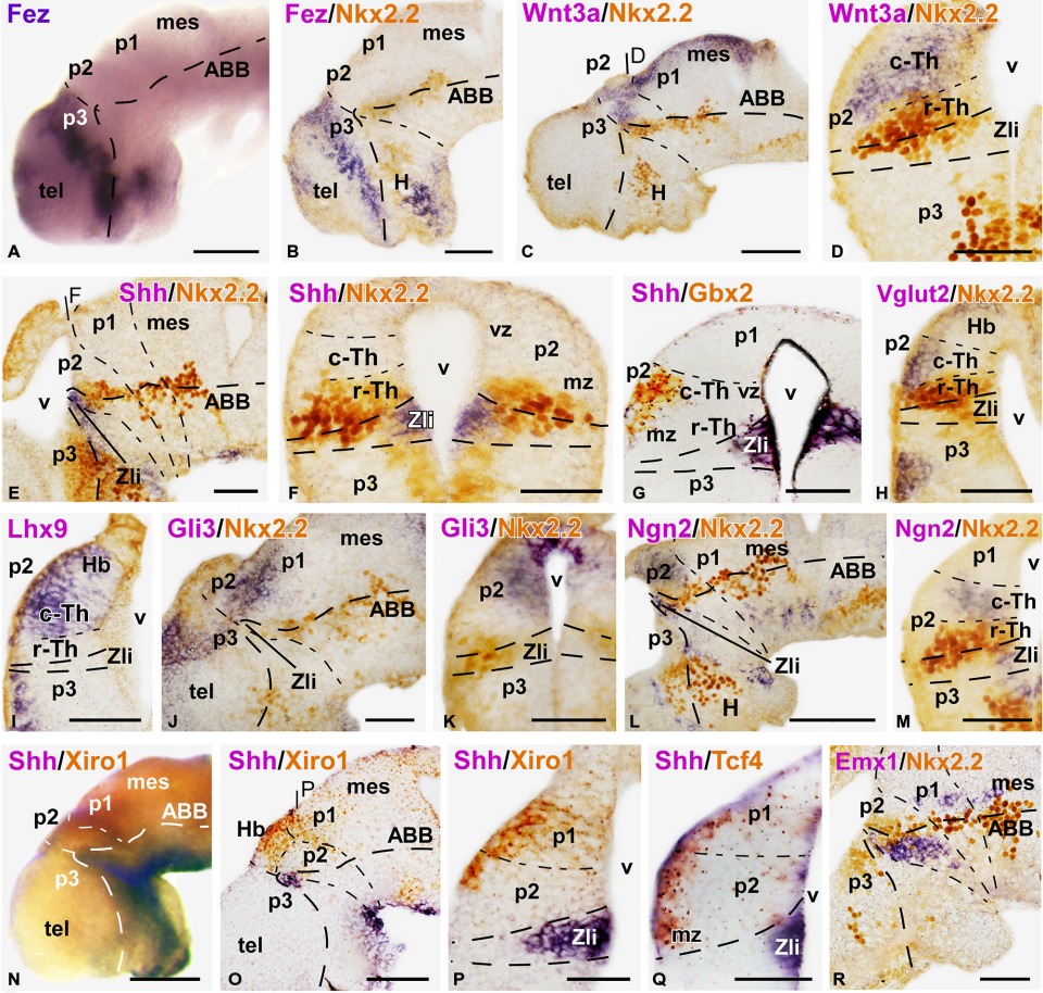
FIGURE 2 | Expression of thalamic markers at early embryonic stages 37/38. Microphotographs of whole mounts (A,N) and sagittal (B,C,E,J,L,O,R) or transverse (D,FâI,K,M,P,Q) sections of embryos at stages 37/38. Photographs correspond to single ISH (purple; A,I), double ISH (purple/orange; G,NâQ) and combination of ISH (purple) with IHC (brown) (BâE, F,H,J,K,L,M,R). The markers labeled are indicated in the upper left of each photograph. All images are oriented following the same standard: dorsal is upwards in transverse and sagittal sections, and rostral is to the left in sagittal sections. The neuromeric boundaries and main brain subdivisions are indicated to assist in the precise localization of the labeling. At these stage c-Th and r-Th subdivision of the thalamus were distinguished (M). The levels of the transverse sections (D,F,P) are indicated in photographs (C,E,O), respectively. Scale bars = 100 µm (A,B,L,N,O), 50 µm (CâK,M,PâR). See list for abbreviations.
Image published in: Bandín S et al. (2015)
Copyright © 2015 Bandín, Morona and González. Creative Commons Attribution license
| Gene | Synonyms | Species | Stage(s) | Tissue |
|---|---|---|---|---|
| fezf1.S | fez, fezf | X. laevis | Throughout NF stage 37 and 38 | forebrain prosomere telencephalon zona limitans intrathalamica |
Image source: Published
Permanent Image Page
Printer Friendly View
XB-IMG-146287