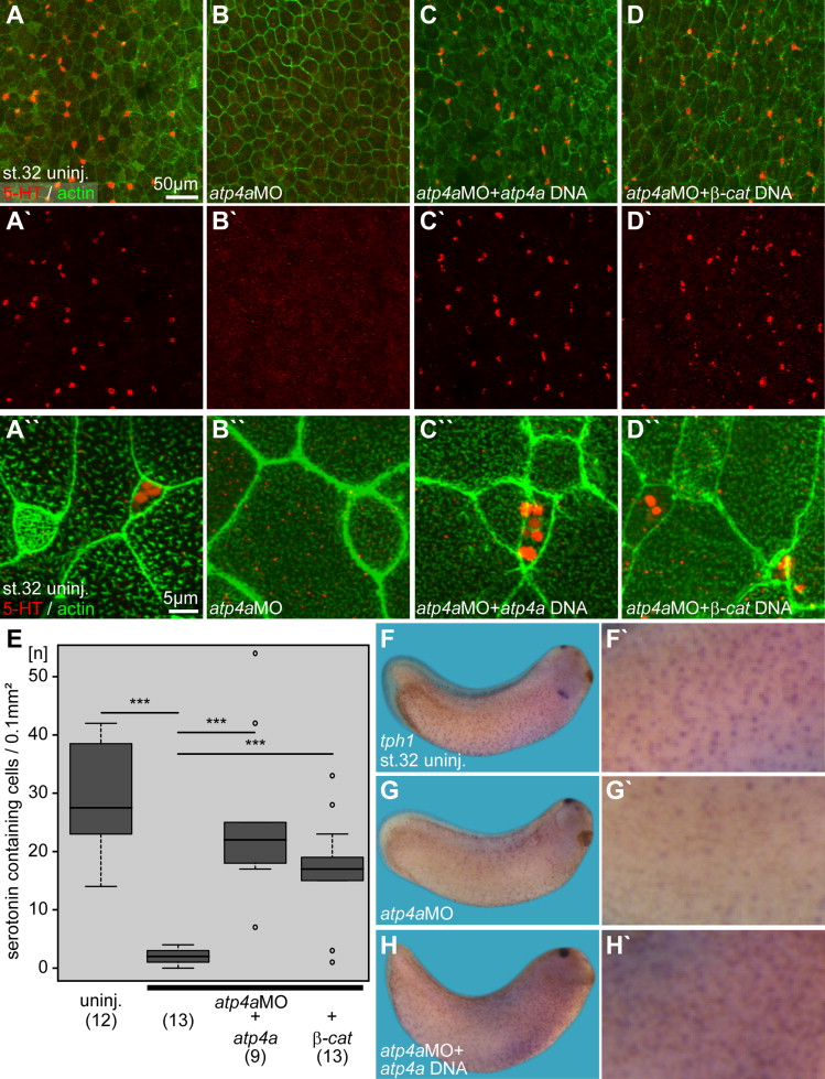
Fig. 5. Serotonin secretion and tph1 expression in the embryonic epidermis are regulated by ATP4a-mediated Wnt/β-catenin signaling. (AâD) Immunofluorescent analysis and quantification (E) of serotonin (5-HT; red) deposition in small secretory cells (SSCs) of the skin at stage 32. Actin (phalloidin-Alexa488) staining in green. (A) Uninjected (uninj.) control. (B) Loss of serotonin staining in atp4a morphants. (C,D) Rescue of serotonin staining upon co-injection of atp4a (C) or β-catenin (β-cat.; D) DNA constructs. (AâµâDâµ) Serotonin channel. (Aâ¶âDâ¶) Higher magnification of (AâD). (E) Quantification of results. (FâH) WMISH for tph1. (F, Fâµ) Uninjected control. (G, Gâµ) Loss of tph1 expression in atp4a morphants was rescued by co-injection of atp4a DNA (H) (n, number of embryos; st., stage).
Image published in: Walentek P et al. (2015)
Copyright © 2015. Image reproduced with permission of the Publisher, Elsevier B. V.
| Gene | Synonyms | Species | Stage(s) | Tissue |
|---|---|---|---|---|
| tph1.L | X. laevis | Throughout NF stage 32 | epidermis ciliated epidermal cell foregut midbrain pineal gland |
Image source: Published
| Experiment + Assay | Source | Phenotypes and Disease | ||||
|---|---|---|---|---|---|---|
| Xla Wt + atp4a MO + NF32 (in situ hybridization) | Fig. 5 G G' |
|
Permanent Image Page
Printer Friendly View
XB-IMG-144159