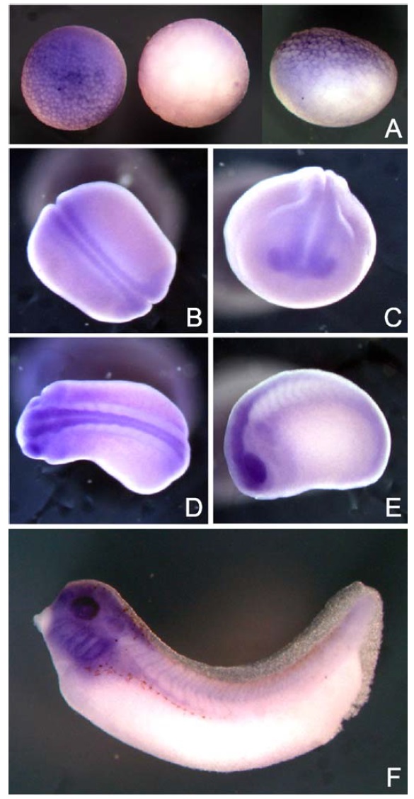
Fig. 2. Spatial expression patterns of xMBD3 transcripts in Xenopus embryos on whole-mount in situ hybridization. (A) Animal pole (left), vegetal pole (middle), and lateral (right) views at stage 8. Dorsal (B) and anterior (C) views of neural groove embryos at stage 19. Dorsal (D) and lateral (E) views of tailbud embryos at stage 23. (F) Lateral view of a tadpole stage embryo.
Image published in: Iwano H et al. (2004)
Copyright © 2004. Image reproduced with permission of the Publisher, Elsevier B. V.
| Gene | Synonyms | Species | Stage(s) | Tissue |
|---|---|---|---|---|
| mbd3.S | LOC108712329, MGC69548, xmbd3 | X. laevis | Throughout NF stage 19 | optic vesicle neural plate pre-chordal neural plate chordal neural plate |
| mbd3.S | LOC108712329, MGC69548, xmbd3 | X. laevis | Throughout NF stage 23 | eye optic vesicle cranial neural crest head neural tube posterior neural tube anterior neural tube |
| mbd3.S | LOC108712329, MGC69548, xmbd3 | X. laevis | Throughout NF stage 33 and 34 | eye pharyngeal arch mandibular arch hyoid arch branchial arch brain forebrain midbrain hindbrain otic vesicle |
| mbd3.S | LOC108712329, MGC69548, xmbd3 | X. laevis | Throughout NF stage 8 | animal hemisphere animal |
Image source: Published
Permanent Image Page
Printer Friendly View
XB-IMG-135547