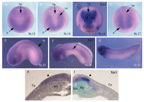
Figure 1. Expression of skp2 and Xic1. Whole-mount in situ hybridisation at indicated stages show expression of (a,b,d,e,g) skp2 and (c,f) Xic1. (a,c) Dorsal view with anterior toward the bottom. (b,d) Anterior view with dorsal toward the top. (e-g) Lateral view with anterior to the left. (h,i) Expression of skp2 and Xic1, respectively, in a vibratome section of stage 16 embryos; primary neurons are indicated with black arrows. Ep, epidermis; I, intermediate stripe; L, lateral stripe; M, medial stripe; My, myotome; Nf, neural folds; Not, notochord; Np, neural plate; Nt, neural tube; P, placodes.
Image published in: Boix-Perales H et al. (2007)
Copyright © 2007 Boix-Perales et al. Creative Commons Attribution license
| Gene | Synonyms | Species | Stage(s) | Tissue |
|---|---|---|---|---|
| skp2.L | xskp2 | X. laevis | Throughout NF stage 13 to NF stage 14 | preplacodal ectoderm anterior placodal area |
| cdknx.L | p27Xic-1, p27XIC1, p28, Xic-1, Xic1 | X. laevis | Throughout NF stage 14 | neural plate |
| skp2.L | xskp2 | X. laevis | Throughout NF stage 17 | neural plate optic vesicle |
| skp2.L | xskp2 | X. laevis | Throughout NF stage 25 | optic vesicle neural tube neural crest cranial neural crest |
| cdknx.L | p27Xic-1, p27XIC1, p28, Xic-1, Xic1 | X. laevis | Throughout NF stage 26 | myotome neural tube |
| skp2.L | xskp2 | X. laevis | Throughout NF stage 33 and 34 | eye brain central nervous system spinal cord pharyngeal arch mandibular arch hyoid arch branchial arch |
Image source: Published
Permanent Image Page
Printer Friendly View
XB-IMG-122555