XB-IMG-205291
Xenbase Image ID: 205291
|
||||||||||
|
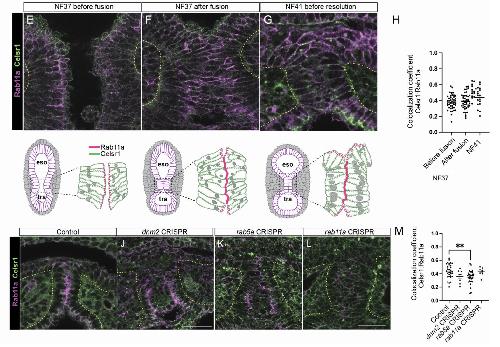
Figure S5. Celsr1 is localized to the periphery of cells during tracheoesophageal separation, related to Figure 6. [continued panels e-M]
S5E-H: Time course of Celsr1 immunostaining during Xenopus trachea-esophageal morphogenesis. Diagrams depict Celsr1/Rab11a localization during foregut separation.
S5I-M: Celsr1:Rab11a colocalization is significantly decreased in rab5a Xenopus mutants, but Celsr1 cellular localization is
not significantly altered in dnm2, rab5a, and rab11a Xenopus mutants (mean min/max, 1W-ANOVA, **p<0.01. n=3-5 cells,
5 embryos analyzed).
Image published in: Edwards NA et al. (2025) Copyright © 2025. This is an open access article distributed under the terms of the Creative Commons CC-BY 4.0 license, which permits unrestricted use, distribution, and reproduction in any medium, provided the original work is properly cited.
Image source: Published Larger Image Printer Friendly View |