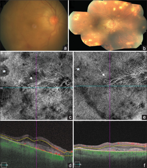
Figure 1. Fundus photograph of the right eye with active inflammation in VKH and peripapillary, CNV (a); sunset glow fundus with peripheral retinal pigment epithelial defects in the right eye with peripapillary iCNV extending to become sub foveal (b); OCTA 6 × 6, en face of the filamentous iCNV (c); flow void areas suggestive of granulomas (white star) in the outer retina-choriocapillaris slab, in the active inflammatory stage and hypo reflective zone around the iCNV marked with white arrows with flow signal in the iCNV (d); OCTA, en face, showing resolving iCNV with clearer hyper reflective vessels (e); decreased hypo reflective zone around the iCNV (white arrow) and decreased flow voids of the granuloma (white star) and corresponding cross-section of the iCNV with compact SHRM and reduced flow signal after intravitreal anti-VEGF (f)
Image published in: Rao V et al. (2026)
Image downloaded from an Open Access article in PubMed Central. Copyright: © 2026 Indian Journal of Ophthalmology
Permanent Image Page
Printer Friendly View
ECB-IMG-208674