XB-IMG-153778
Xenbase Image ID: 153778
|
|||||||||||||||
|
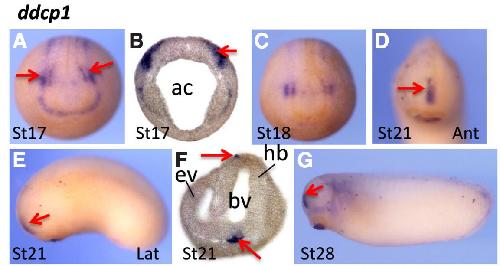
Fig. 2. Spatial expression pattern of ddcp1 encoding a Duf domain protein.
(A-C) ddcp1 was expressed along the edge of neural plate. Notably, two stronger
staining patches (arrows) were detected in whole mount embryos (A) and transverse
sections (B). With the closure of neural tube, the two signaling patches merged
and were located at the frontal region of the head (D-G). Ant, anterior view; lat,
lateral view. Ac, archerteron; ev, eye vesicle; bv, brain vesicle; hb, hindbrain. Image published in: Wong TC et al. (2016) Copyright © 2016. Image reproduced with permission of the Publisher, University of the Basque Country Press.
Image source: Published Larger Image Printer Friendly View |