XB-IMG-149072
Xenbase Image ID: 149072
|
||||||||||
|
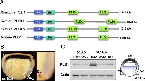
Fig. 1. PLD1 is dynamically expressed during Xenopus development. (A) Schematic diagrams of PLD1 in various species. PLD1 is well conserved among vertebrates. The number of amino acids in each PLD1 protein is indicated. (B) In situ hybridization assay with PLD1 and sagittal section views. Endogenous PLD1 was expressed in the involuting mesoderm and the overlying ectoderm. A white arrow indicates dorsal lips of stage 10.5 embryos and right panel is a magnified view of a white rectangle. (C) PLD1 protein was asymmetrically enriched in the dorsal region. Dorsal Marginal Zone (DMZ), Ventral Marginal Zone (VMZ), and Animal Cap (AC) were subjected to western blotting with anti-PLD1 antibody. Image published in: Lee H et al. (2016) Copyright © 2016. Image reproduced with permission of the Publisher, Elsevier B. V.
Image source: Published Larger Image Printer Friendly View |